Cranial Nerve VII: Facial Nerve
What’s unique about the Facial Nerve?
The facial nerve is a complex nerve with motor, sensory, and parasympathetic functions. It travels a long intraosseous route, taking several turns before emerging out of the base of the temporal bone.
What is the function of the nerve?
Motor: Muscles of facial expression (stapedius, stylohyoid, and posterior belly of digastric)
Sensory: Sense of taste (anterior 2/3 of the tongue)
Parasympathetic: Lacrimal gland (tear production), sublingual and submandibular glands (saliva production), as well as aspects of the mucosa of the nose, pharynx, and palate.
What are the signs of dysfunction?
Symptoms of facial nerve paralysis vary depending on the cause:
• Unclear speech or slurred speech
• Drooling and challenges eating and drinking
• Drooping eyebrow on the affected side of the face
• Facial twitches (tics)
• Inability to move facial muscles such as your forehead, eyebrow, and corner of your mouth
• Asymmetrical smile or facial appearance
• Loss of smell or taste
• Nasal blockages
• Trouble closing your eyes or blinking
• Amplified sounds from your ear on the affected side of the face
How might this nerve be impacted?
Several conditions can cause weakness or paralysis of the facial nerve:
• Accidents and facial fractures
• Bell’s palsy (inflammation of the facial nerve)
• Cancer such as salivary gland cancer and meningioma (skull base tumor)
• Ear infections or ear tumors like acoustic neuromas and schwannomas
• Facial surgery
• Guillain-Barré syndrome, an autoimmune disease
• Lyme disease
• Nerve blocks
• Ramsay Hunt syndrome (neurological disorder caused by infection from chickenpox or shingles virus)
• Sarcoidosis
• Stroke
How can you work with this nerve?
• Notice the temporal bones and offer space for decompression
• Visualize the facial nerve pathway, from the brainstem, through the temporal bone, and out to the 5 branches
• Notice the quality of potency moving through the nerve
• Hold space for any held patterns along the pathway to shift
The facial nerve emerges from the lower margin of the pons where it meets the medulla oblongata.

It emerges as two distinct routes- one of which is the motor route, often called the facial motor route (or the facial nerve proper), which contains motor neurons.

The second route is the intermediate nerve, which carries sensory and parasympathetic fibers.

These two nerves remain distinct as they enter the internal acoustic meatus in the petrous part of the temporal bone. They merge just inside the bone.

Just inside the temporal bone is the geniculate ganglion, located on the first of three 90-degree turns of the facial nerve.

The greater petrosal nerve branches from the ganglion inside the bone and travels anteriorly to the pterygopalatine ganglion to supply the lacrimal, nasal, palatine, and pharyngeal glands with parasympathetic innervation.

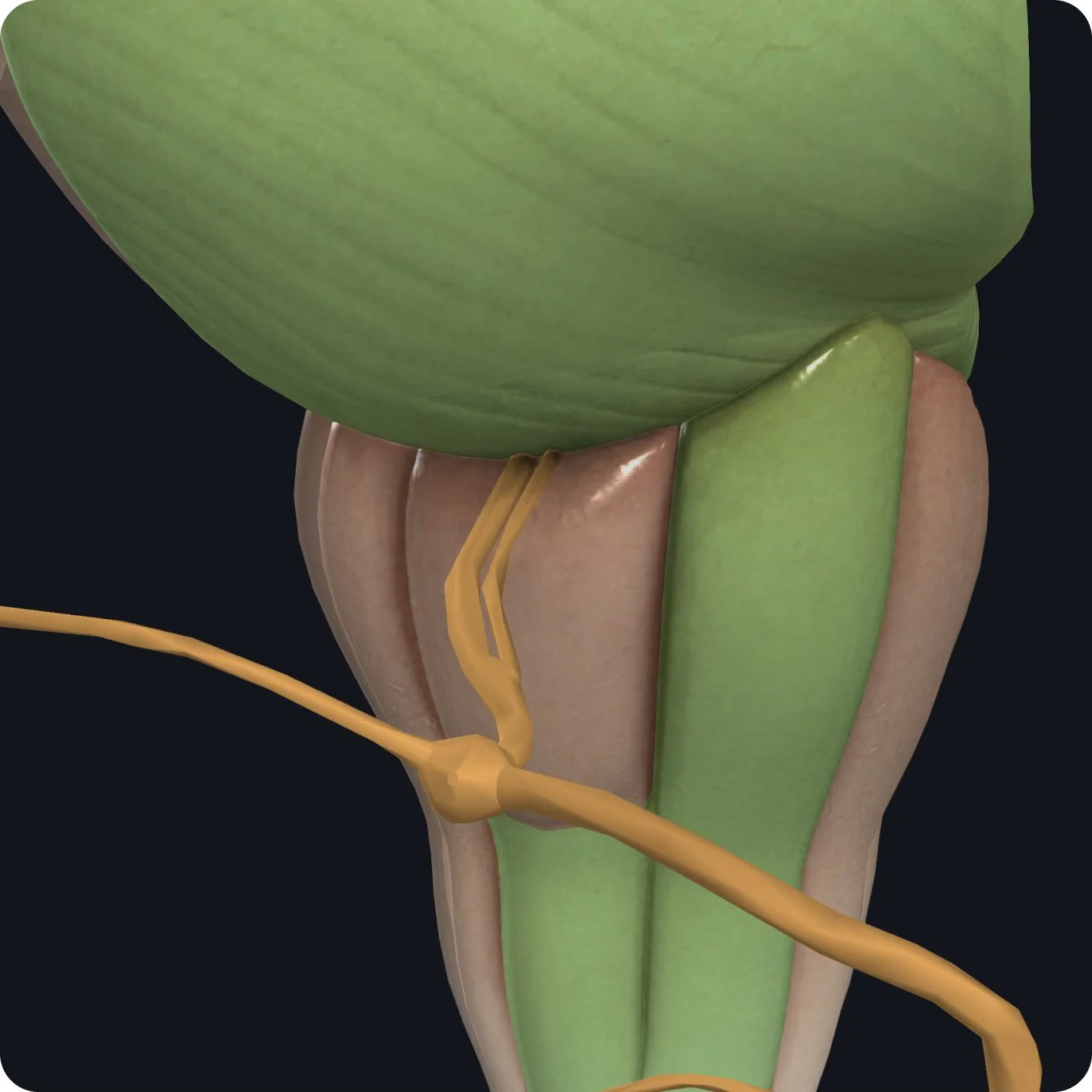
Inside the bone, just after the second 90-degree turn, a small branch passes anteriorly into the middle ear; this is chorda tympani. It runs behind the tympanic membrane.

It exits on the anterior side of the temporal bone to join the lingual nerve where it picks up special sensory information from the anterior 2/3 of the tongue. Chorda tympani carries parasympathetic innervation to the submandibular and sublingual glands via the lingual nerve.

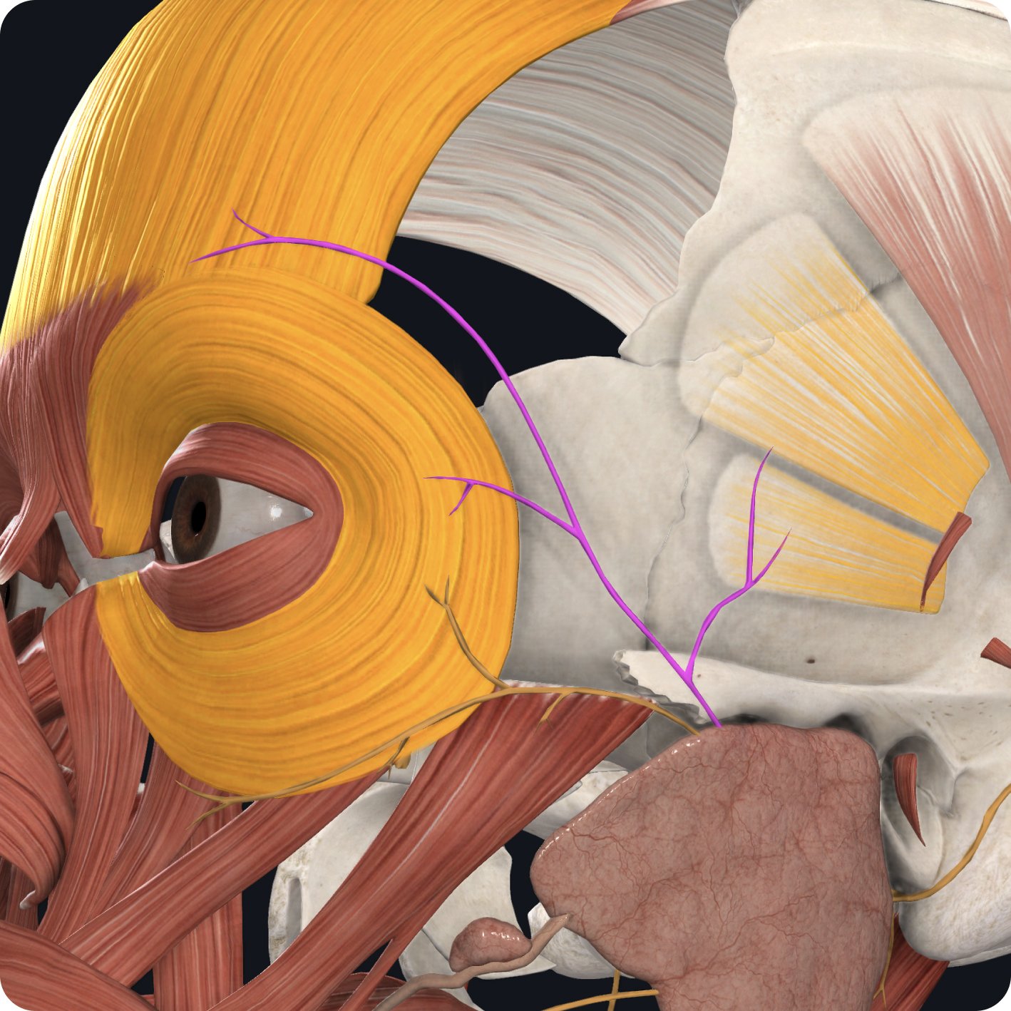
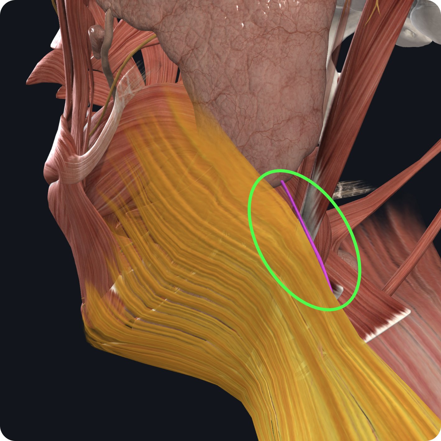
The facial nerve takes one last intracranial turn before it emerges from the stylomastoid foramen, a small hole located between the styloid process and the mastoid process of the temporal bone. It's not visible here, but before it emerges the facial nerve gives a small branch to the stapedius muscle within the middle ear.

Following its exit from the cranium, two small sylohyoid branches emerge medially to provide innervation to the stylohyoid muscle.

Laterally, a digastric branch innervates the digastric muscle.

The posterior auricular nerve is a larger motor branch that bifurcates from the terminal branch and passes posteriorly.supplying the occipital belly of occipitofrontalis and arecularis muscles.

The terminal part of the facial nerve enters the posterior edge of the parotid gland, dividing into five branches.
The temporal branch supplies muscles of facial expression around the upper part of the face- including the eye, eyebrows, forehead, and ear.

The zygomatic branch supplies muscles of facial expression around the orbit and upper lip.

The buccal branch supplies muscles of facial expression around the middle of the face, cheeks, and mouth.

The mandibular branch supplies muscles of facial expression around the lower face and jaw.
The cervical branch of the nerve supplies the platysma muscle of the neck.