Cranial Nerve VIII: Vestibulocochlear Nerve
What’s unique about the Vestibulocochlear Nerve?
The vestibulocochlear nerve courses in parallel to the facial nerve. As its name implies, the vestibulocochlear nerve has two divisions; the “vestibular” division provides our sense of balance, and the “cochlear” division provides our sense of hearing.
What is the function of the nerve?
Sensory: Sense of hearing and balance
What are the signs of dysfunction?
Signs of vestibulocochlear nerve dysfunction may include:
• Hearing loss
• Tinnitus, a ringing or hissing sound in the ears
• Vertigo, which presents either subjectively, where the client senses that they are moving, or it can be objective where the client feels that objects in their environment are moving
• Balance challenges
• Nystagmus (involuntary movement of the eyes)
How might this nerve be impacted?
Damage to the nerve can occur due to:
• Trauma
• Viral infection within the inner ear, in the area surrounding the nerve, or elsewhere in the body
• Congenital differences
• Tumor
• Vascular injury (injury to an artery or vein)
• Compression of the brainstem or other surrounding structures
How can you work with this nerve?
• Notice the brainstem and temporal bones. Notice if there is a request for decompression in either or both places, and offer that if so. Otherwise, offer decompression along the nerve pathway.
• Visualize the pathway, from between the pons and medulla, moving alongside the facial nerve to enter the temporal bone through the internal acoustic meatus, splitting into vestibular and cochlear divisions to access the vestibular organ and cochlea.
• Notice the quality of potency moving through the nerve.
• Hold space for any held patterns to shift.

The vestibulocochlear nerve first emerges from the brainstem between the pons and medulla.

The Vestibulocochlear nerve courses in close association with the facial nerve.
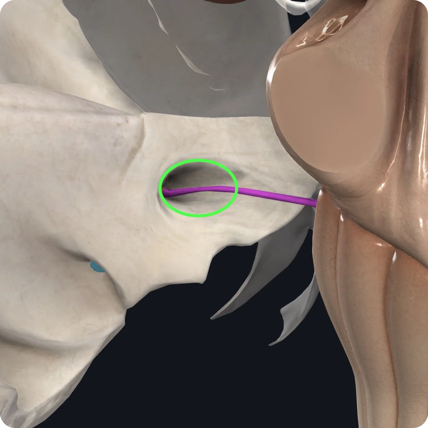
It then enters the petrous part of the temporal bone through the internal acoustic meatus.

The nerve divides into an anterior trunk, the cochlear nerve, and a posterior trunk, or the vestibular nerve.
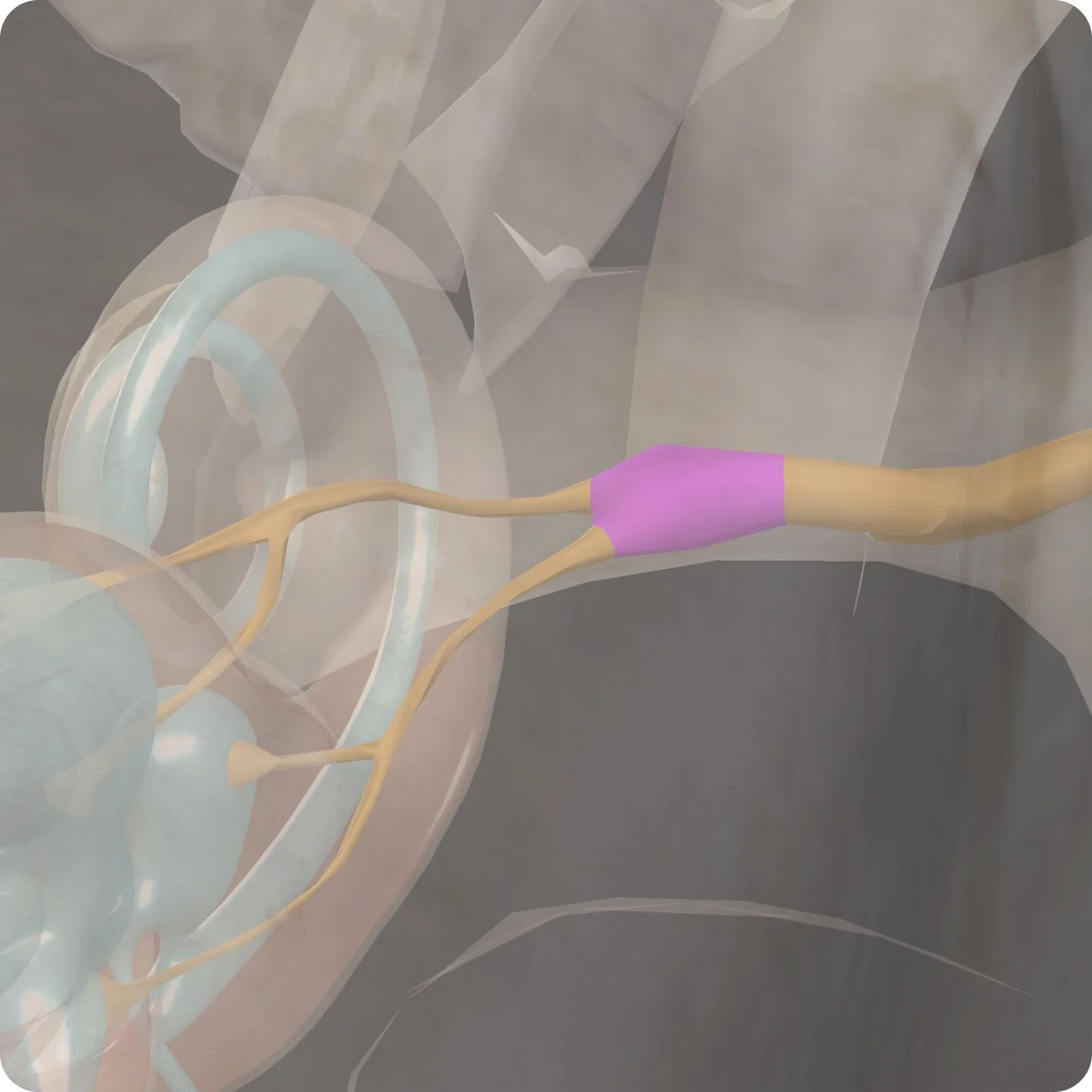
After the division of the nerve, the vestibular nerve passes to the vestibular ganglion, a collection of sensory neuronal cell bodies and divides into superior and inferior branches.

The superior branch conveys sensory information from the anterior and lateral semicircular ducts and the Utica with the vestibular organ.
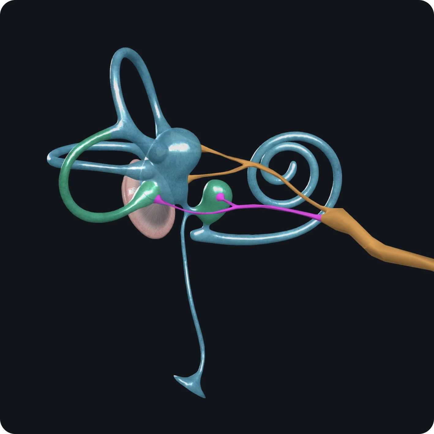
The inferior branch innervates the posterior semicircular duct and the saccule within the vestibular organ.
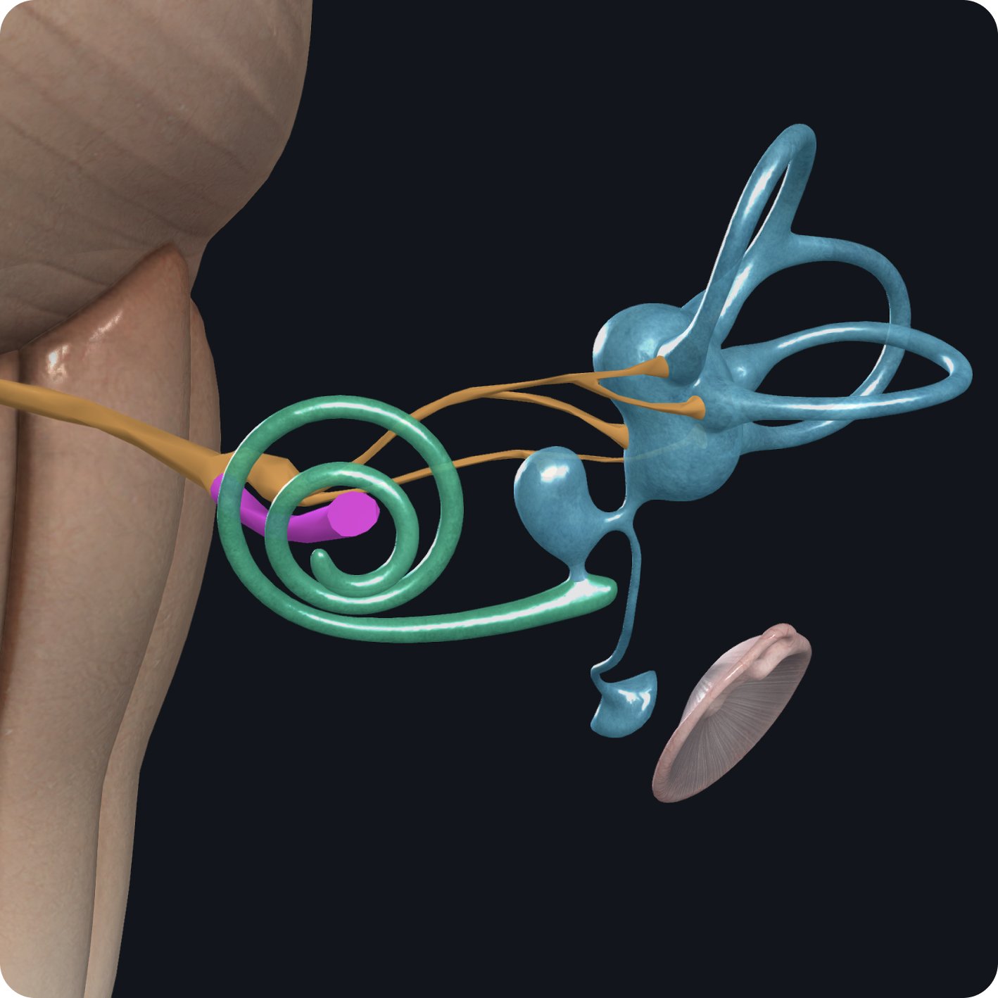
The cochlear nerve travels towards the cochlea and coils up on itself to form a spiral shape within the cochlear duct.

The cochlear nerve makes contact with special hair-like cells in the cochlea.

The hair cells transfer auditory information from movement of the fluid within the cochlea through the cochlear nerve to the auditory cortex of the brain, enabling hearing.

The vestibular part of the nerve is responsible for maintaining posture and balance through rotational movement and processing the position of the head with respect to gravity.