Cranial Nerve V: Trigeminal Nerve
What’s unique about the Trigeminal Nerve?
The Trigeminal nerve has both motor and sensory functions “Tri” means three, and the Trigeminal nerve has three major divisions. It is responsible for sending pain, touch, and temperature sensations from the face to the brain. The mandibular section of the nerve supports chewing and swallowing.
What is the function of the nerve?
Sensory: Sensation to the face, surface of the eye, nasal and oral cavities, and general sense to the anterior 2/3 of the tongue (sensing temperature and texture, but not taste)
Motor: Muscles of mastication (chewing)
What are the signs of dysfunction?
Signs of damage to the Trigeminal nerve may include:
• Difficulty chewing or speaking
• Ongoing numbness or facial pain in the area that the damaged nerve senses
• Sudden, intense facial pain on one side of your face
• Chronic aching or burning feeling on the face
A Trigeminal nerve injury may affect a small area, like part of your gum, or a large area, like one side of your face.
How might this nerve be impacted?
Sometimes people can present with Trigeminal neuralgia, brought on by nerve damage.
Damage to the nerve can occur due to:
• An artery or nerve wrapping around the Trigeminal nerve
• A tumor or cyst creating pressure on the nerve
• Traumatic injury to the face
• Infection residing in the nerve may only affect one division (For example, herpes zoster more commonly known as facial shingles).
How can you work with this nerve?
• Notice the brainstem and pons, the periosteal and meningeal dura, the temporal bone, and the face field. Offer space for decompression for each.
• Visualize the pathway, sensory and motor roots arising from the side of the pons, the presence of the trigeminal ganglion surrounded by the dura alongside the cavernous sinus, and the splitting into the 3 branches into the forehead, face and cheek, and jaw.
• Notice the quality of potency moving through the nerve.
• Hold space for any held patterns to shift.
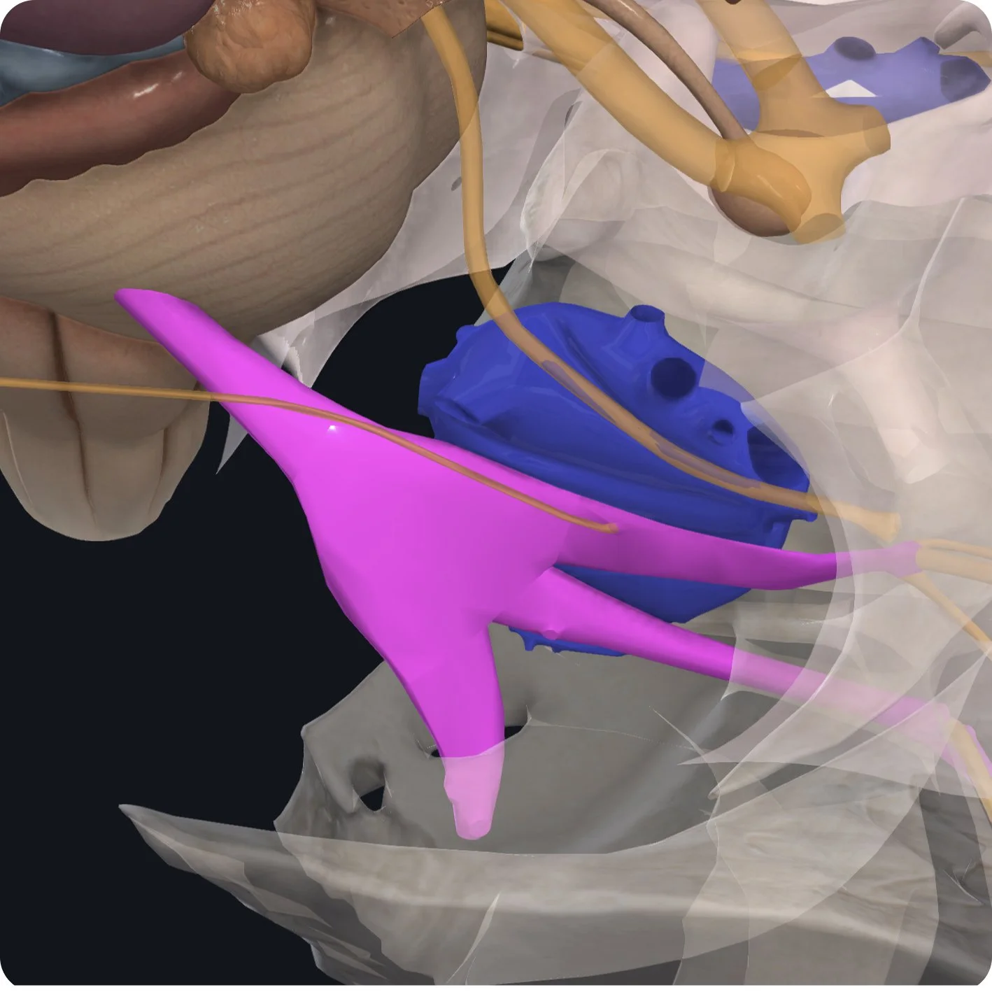
The Trigeminal nerve emerges from the brainstem on the lateral aspect of the pons.

It has two distinct roots. The sensory root is the larger of the two and carries sensory information into the brainstem.

The motor root carries motor information into the periphery.

Both roots come together at the trigeminal ganglion.

This flat, crescent-shaped structure lies on the floor of the temporal bone, between the periosteal dura and meningeal dura.

It has its own expansion of meningeal dura called meckel's cave. The unusual shape of the trigeminal ganglion is due to the large number of sensory nerve cell bodies that are within it.

The trigeminal ganglion receives information from three branches. The ophthalmic branch superiorly carries sensory information towards the forehead (V1).

The maxillary branch in the middle also carries sensory information towards the face and cheek (V2).

Inferiorly, the mandibular branch carries both motor and sensory information towards the jaw (V3).