Cranial Nerve II: Optic Nerve
What’s unique about the Optic Nerve?
The Optic Nerve has a purely sensory function, providing our sense of sight. It transmits visual fibers from our retina and sends them back to the brain. Some of this information crosses from one side to the other at the optic chiasm.
What is the function of the nerve?
Sensory: Sense of vision
What are the signs of dysfunction?
Signs of optic nerve dysfunction vary:
• Color blindness
• Eye pain
• Headaches
• Nausea and vomiting
• Night blindness
• Partial or complete vision loss
• Peripheral (side) vision loss
• Ringing in your ears (tinnitus)
How might this nerve be impacted?
These conditions can damage an optic nerve and affect vision:
• Glaucoma
• Anterior ischemic optic neuropathy
• Congenital abnormalities
• Optic atrophy
• Optic nerve coloboma
• Optic nerve drusen
• Optic nerve gliomas
• Optic nerve meningiomas
• Papilledema
• Devic’s disease
How can you work with this nerve?
•Notice the occiput and visual cortex, the sphenoid bone, and the eye field and offer space for decompression for each
• Visualize the optic pathway, from the eyes, moving through the optic canal of the sphenoid, crossing at the chiasm, extending into optic tracts, and traveling through the lateral geniculate nucleus of the thalamus
• Notice the quality of potency moving through the nerve, and sense right vs. left
• Hold space for any held patterns to shift
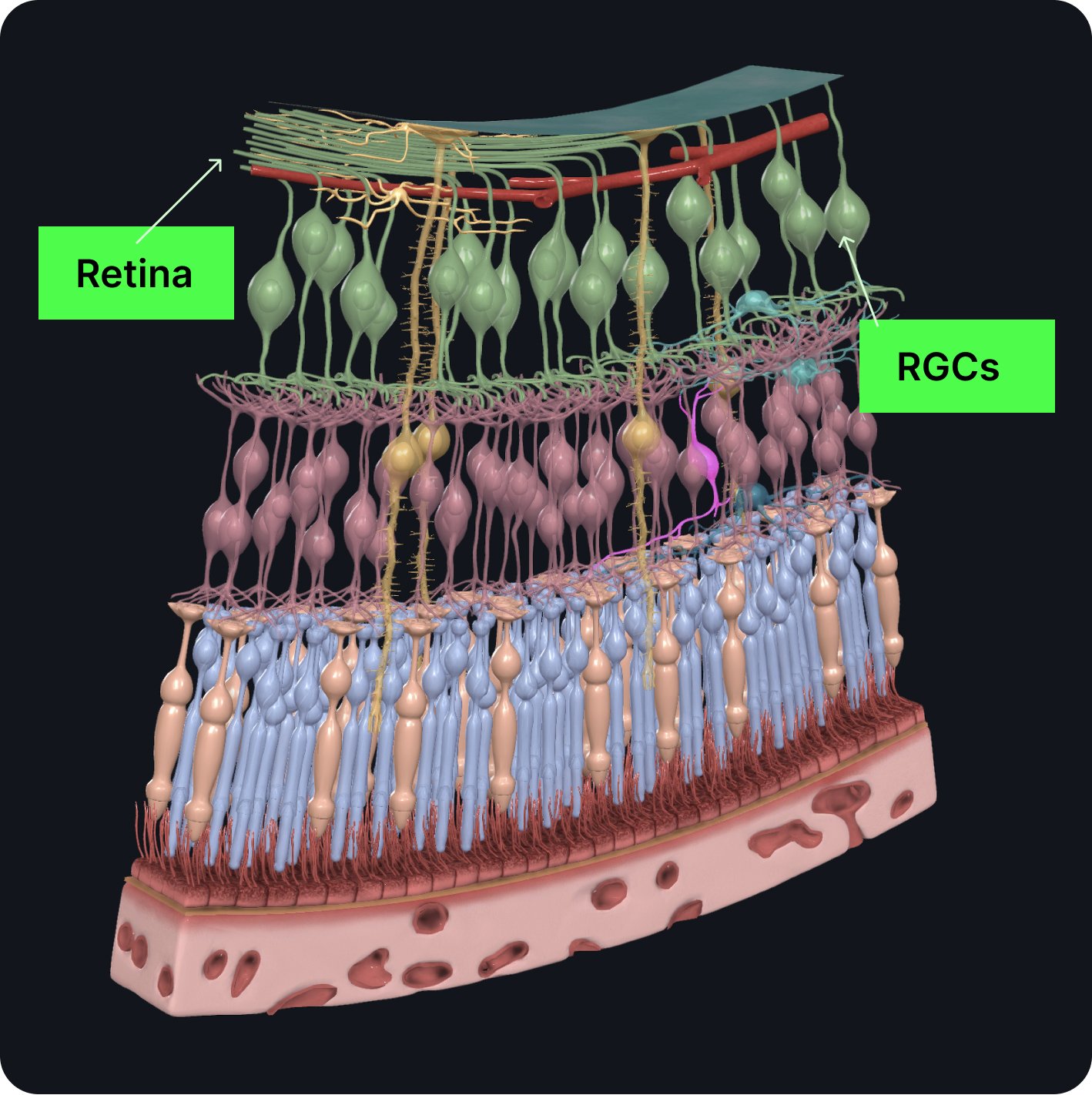
The journey of the optic nerve begins at the retina which is formed by the convergence of axons from the retinal ganglion cells (RGCs).

These cells in turn receive impulses from the photoreceptors of the eye, known as the rods and cones.

After its formation, the optic nerve leaves the bony orbit via the optic canal, a passageway through the sphenoid bone.

It enters a cranial cavity running through the chiasmatic sulcus in close proximity to the pituitary gland.

Within the middle cranial fossa the optic nerves from each eye unite to form the optic chiasm.

The optic chiasm then continues on to form the optic tract.

Each optic tract travels to the ipsilateral (same side) cerebral Hemisphere via the lateral geniculate nucleus, a relay system in the thalamus.

Both the visual field and the retina of each eye is divided into nasal and temporal halves. Information from the visual field is perceived in the opposite side of the retina.

At the optic chiasm, fibers from the nasal half of each retina cross over to the opposite side. Fibers from the temporal halves remain on the same side A combination of fibers from the nasal and temporal halves of each retina forms the optic tract.