Cranial Nerve VI: Abducens Nerve
What’s unique about the Abducens Nerve?
The abducens nerve has motor-only functionality and is one of the most straight-forward of the cranial nerves. It is responsible for moving 1 out of the 6 extraocular muscles, the lateral rectus, which “abducts” the eye, moving it outward and away from the midline.
What is the function of the nerve?
Motor: Innervation of the lateral rectus muscle to create lateral movement of the eye (moving the eye away from the midline, or abducted)
What are the signs of dysfunction?
The abducens nerve is thin and delicate, and its course is complex. It is therefore especially vulnerable to damage.
Damage to the abducens nerve, often named cranial nerve VI palsy, may lead to the eye drifting medially and inward towards the midline. This symptom may be intermittent or persistent.
How might this nerve be impacted?
Damage to the nerve can occur due to:
• Trauma
• Compression via cavernous sinus thrombosis or internal carotid artery aneurysm
• Hydrocephalus, buildup of fluids in the ventricles
• Infection
• Inflammation
• Intracranial tumor
• High or low intracranial pressure, often associated with headache or drowsiness
• Meningitis
• Microvascualar ischemia
• MS
• Stroke
• Congenital or birth impacts
How can you work with this nerve?
• Notice the ventricles. Notice if there is a request for decompression and drainage, and offer that if so. Otherwise, offer decompression along the nerve pathway and its nearby structures.
• Visualize the pathway, from the base of the pons, crossing the clivus of the occiput, through the tentorium, moving along the petrous part of the temporal bone, moving through the cavernous sinus along the internal carotid artery, and moving through the superior orbital fissure of the sphenoid.
• Notice the quality of potency moving through the nerve.
• Hold space for any held patterns to shift.

The abducens nerve arises from the abducens nucleus in the pons of the brainstem. The axons of motor neurons emerge from the brainstem between the pons and the medulla oblongata.

The nerve then runs forward and superiorly along the surface of the clivus within the subarachnoid space.

It pierces the dura as it arches forward.

It then positions itself directly over the petrous part of the temporal bone.

It passes through the medial wall of the inferior petrosal sinus, and runs into the cavernous sinus.
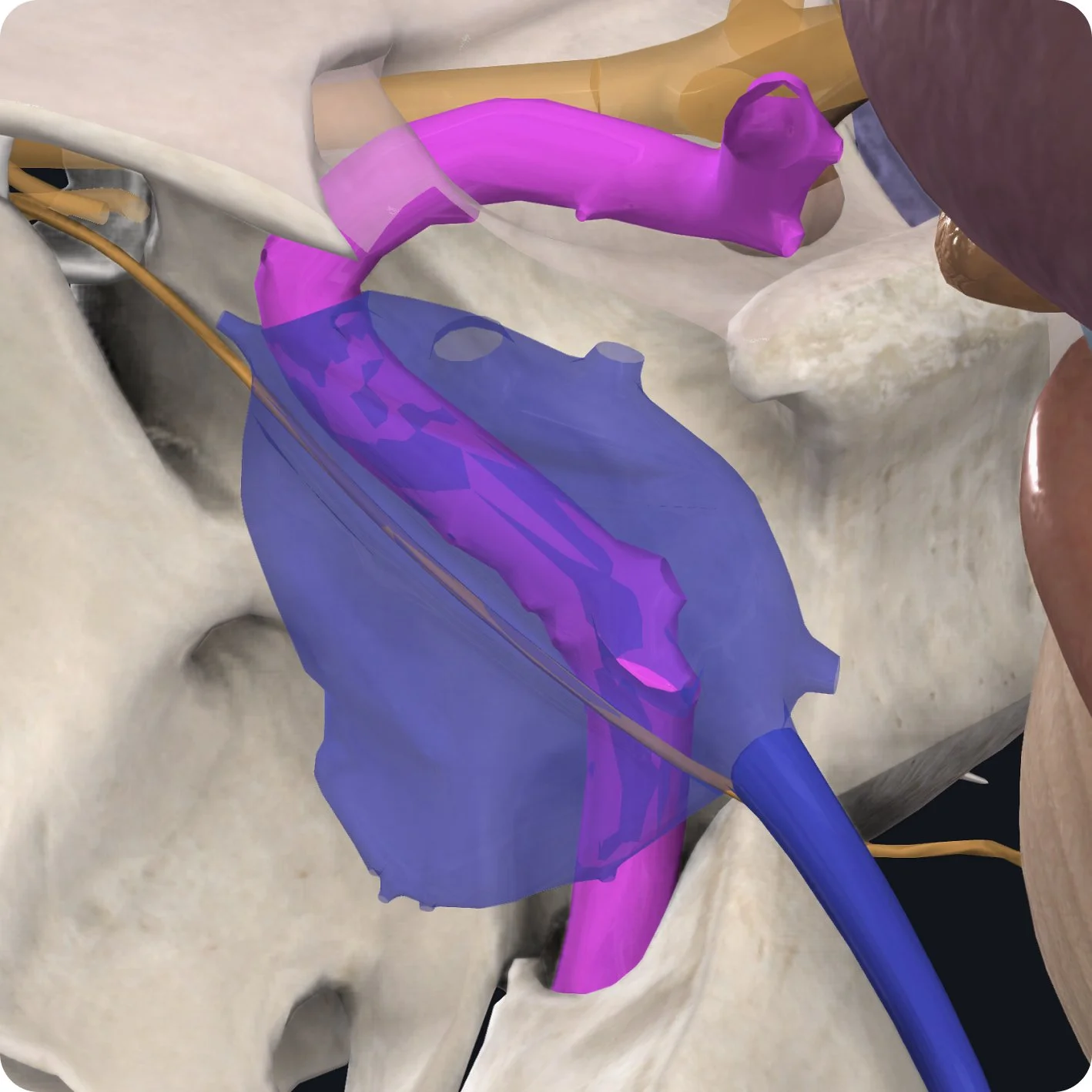
In the cavernous sinus the abducens nerve lies lateral to the internal carotid artery.

Upon exiting the sinus, the abducens nerve leaves the cranium via the superior orbital fissure within the common tendinous ring.

Within the bony orbit, the abducens nerve then terminates by innervating the medial surface of the lateral rectus muscle.

The lateral rectus muscle supports lateral movement of the eyeball, which moves the eye away from the midline (abducted).